Weekly Brain Slice: Superior Colliculus
- Pamela Brown

- Nov 25, 2025
- 5 min read
Updated: Dec 7, 2025
A weekly deep dive into the hidden architecture of your mind.
The Superior Colliculus: The Reflex Engine Behind Your Gaze
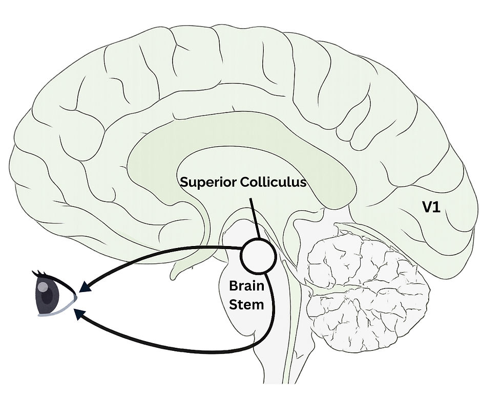
Where It Lives
The superior colliculus (SC) lives in the back of the midbrain. It sits above the inferior colliculus and behind the pineal gland. Together, the superior and inferior colliculi form the tectum (“roof”) of the midbrain.
This location is important because it places the SC right in the path of fast visual and auditory information entering the brain.

What It Does
The superior colliculus receives visual, auditory, and somatosensory inputs and uses them to orient your head and eyes toward meaningful stimuli. In other words, it’s constantly scanning for what matters and guiding your gaze toward it.
How It Works
Just like the LGN, the superior colliculus receives visual signals directly from the retina. 10% of the ganglion cells in the retina project to the SC (often called the retinotectal projection). It also receives auditory input from the inferior colliculus and visual association input from the parietal cortex. Deeper layers integrate tactile, body-position, and sensory context.
This structure tells your eyes where to move so you can center important objects onto your fovea (the part of your retina packed with cones for sharp vision). When something moves in your visual field, the SC is what causes your eyes to immediately jump toward it.
Why the Superior Colliculus Matters
The SC plays a major role in orienting your gaze. Damage to this area makes it harder to look directly at certain stimuli, especially sudden or unexpected ones.
Clinical Connection
The superior colliculus plays a key role in fast eye movements, attention shifts, and automatic responses to visual stimuli. When this system functions differently, these changes can show up clinically:
Parkinson Disease: Slower or harder-to-initiate saccades due to reduced dopamine signaling.
Autism: Reduced automatic orienting to faces or emotional cues in early development.
ADHD: Altered SC–frontal connections may contribute to distractibility and difficulty filtering visual noise.
Migraines (Aura): SC involvement may amplify motion sensitivity and the difficulty focusing during visual aura.
Blindsight: In blindsight, people who are “blind” in part of their vision can still react to movement in that space because the SC pathway is still active.
Lesions/Injury: Slowed gaze shifts and weaker defensive reactions to approaching objects.
Differences in SC pathways can affect how quickly we react, focus, and orient to the world.
Ways to Remember It
If the LGN is your eye's director, think of the SC as your brain’s spotlight operator. It “aims” your eyes toward anything interesting — just like a spotlight operator shining light on the target onstage.
Midbrain = the “middle manager.” It literally sits in the middle of the brain and acts like a manager rapidly directing attention, gaze, and responses.
Layer order = “See → Integrate → Act.” A clean top to bottom flow.
Top layers: visual input
Middle layers: multisensory integration
Bottom layers: motor output
Fun Facts
Superior colliculus is Latin for "little hill".
Even though it only receives 10% of the ganglion cells from the retina, that is still 10,000 neurons, which is equal to the total number of retinal ganglion cells in a cat.
When you instinctively turn your head or eyes toward something in motion, that's the SC activating. It guides your eyes before you are even aware of it.
Babies use it before the visual cortex is mature. Infants track faces and follow gaze early in development thanks to the SC, which is active long before higher cortical pathways come online.
It’s extremely fast. SC circuits trigger motor responses in tens of milliseconds — much faster than conscious visual processing.
It coordinates eye movements and reaching. Deep layers help select the target you look at and reach for, keeping those movements aligned.
Animals rely on it even more than humans. In many species, the SC’s equivalent is their main visual processing center.
Deep Slice:
The superior colliculus technically has seven layers (stacked like a 3D sheet) that fall into three functional zones:
Superficial (Top 3) - Handles pure visual processing and get its input from the retina (mostly contralateral), the primary visual cortex (V1), and other higher visual areas.
1. Stratum zonale – thin top layer receiving broad visual input
2. Superficial gray layer – where retinal and cortical visual signals arrive
3. Optic layer – the main layer receiving retinal axons (important for motion + light detection)
Intermediate (Middle 2) - Integrates multiple sensory signals and gets its input from the inferior colliculus and the auditory cortex
4. Intermediate gray layer – mixes visual + auditory + somatosensory info
5. Intermediate white layer – carries fibers forming early motor commands
Deep (Bottom 2) - Strongly connected to motor pathways and gets its inputs from the primary somatosensory cortex (S1), trigeminal nucleus (facial sensation), and the spinal cord (neck/body position).
6. Deep gray layer – processes looming threats, directs complex orienting responses
7. Deep white layer – sends commands to brainstem and spinal cord motor centers
The neural connections of the superior colliculi are intricate and not yet fully comprehended. These structures primarily receive visual information from the retina through the optic nerve and tract, with less input from the visual cortex and other higher visual areas. Additionally, the superior colliculi receive auditory signals from the inferior colliculi and the auditory cortex to coordinate movement responses with sounds. Further somatosensory projections to the deep layers enable the superior colliculi to react to tactile stimuli.
Basically, the inputs tell the SC what is happening, where it is, and how your body is positioned to respond. As a results the SC sends signals to the neck movement centers, eye-movement centers, and defensive circuits including the PAG and thalamus.
Overview of Key Functions of the SC:
Directing Movements: Turning head and eyes toward (or away from) important stimuli
Saccadic Eye Movements: Initiating quick jumps (saccades) to new targets
There are two different types of saccades - voluntary and reflexive.
Frontal Eye Fields control voluntary and memory-guided saccades, while the SC controls reflexive saccades. However, if the FEF is damaged, the SC can take over after a sort dysfunction period.
Looming Detection: Responding to rapidly approaching objects
Reaching/Arm Movements: Helping select movement targets
Decision-Making: Choosing which stimulus you should orient toward
Face Detection: Early, automatic recognition
Attention Shifts: Redirecting attention even without eye movement
Multisensory Integration: Matching signals from sight, sound, and touch
TL;DR: the SC helps you detect, orient, look, and react.
What I Find Fascinating About It
The SC contains an actual map of your visual world. When an object moves across your vision, the same pattern of movement appears across the SC’s neurons. This map ensures the SC can quickly guide your eyes exactly where they need to move. It contains a literal map of your visual world. Stimuli moving across your visual field cause matched shifts in activity across the SC, almost like a radar sweep.
It’s also excellent at detecting looming threats. The SC can detect a rapidly approaching object, like someone tossing something at your head, and trigger a defensive response (flinch, duck, step back) even if your eyes don’t visibly move. And it does this before you consciously realize what’s happening. It detects threats before you do. Looming objects (like something flying toward your face) activate SC-driven defensive circuits automatically.
Big Picture
The superior colliculi serve as the primary brain regions for integrating both visual and nonvisual information. This multisensory, motor-control center receives input from various sources, including visual, auditory, and tactile stimuli. It constructs a spatial map of the environment and initiates saccadic movements while coordinating the movements of the head, eyes, and arms. Additionally, it responds to approaching threats, links to defensive pathways, and facilitates rapid shifts in attention. Overall, it represents one of the brain's fastest systems for converting sensation into action.
Download the SC Coloring Worksheet:
*Image of relative locations of the M-, P-, and K-layers (macaque monkey) By Pancrat - Own work, CC BY-SA 3.0, https://commons.wikimedia.org/w/index.php?curid=17718719


Comments